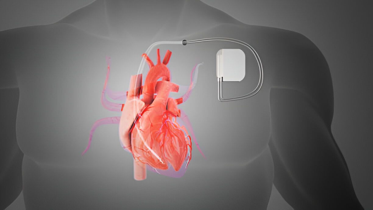
When it comes to heart health, advanced medical procedures like electrophysiology studies are pivotal in diagnosing and treating complex cardiac arrhythmias. At Atrius, we pride ourselves on offering state-of-the-art electrophysiology services to our patients, ensuring they receive the best possible care. In this comprehensive blog, we will delve into what you can expect from advanced electrophysiology procedures at Atrius and why our facility is a trusted name in cardiac care.
Understanding Electrophysiology
Electrophysiology involves the study of the electrical activity of the heart to diagnose and treat abnormal heart rhythms or arrhythmias. This specialised field uses a variety of tests and procedures, including echocardiography.
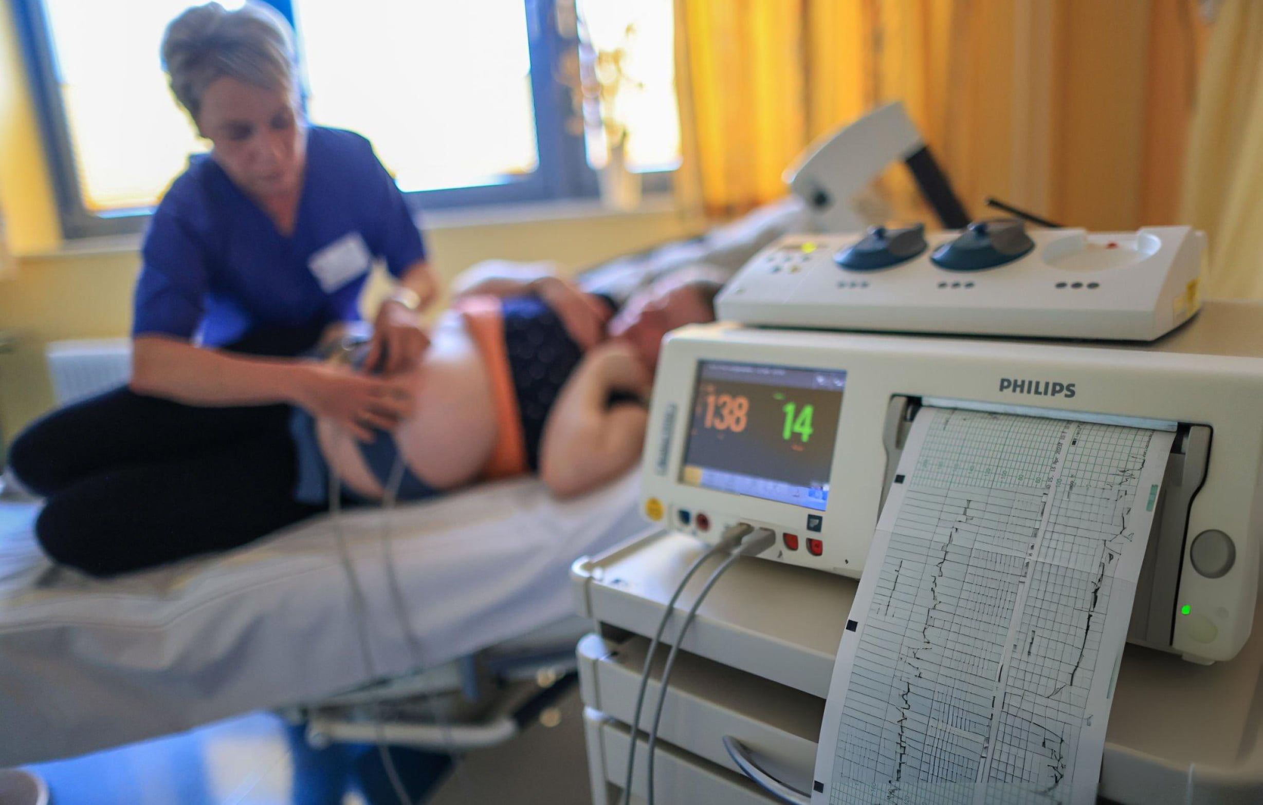
Echocardiography, also known as an echocardiogram, is a type of ultrasound test that uses high-frequency sound waves (echo) to create images of the heart. There are various types of echocardiography, each serving different diagnostic purposes.
Key Components of Echocardiography
- Transthoracic Echocardiography (TTE): The most common type, involving a transducer placed on the chest.
- Transesophageal Echocardiography (TEE): Involves passing a specialised probe through the oesophagus.
- Stress Echocardiography: Performed during or after exercise to evaluate heart function under stress.
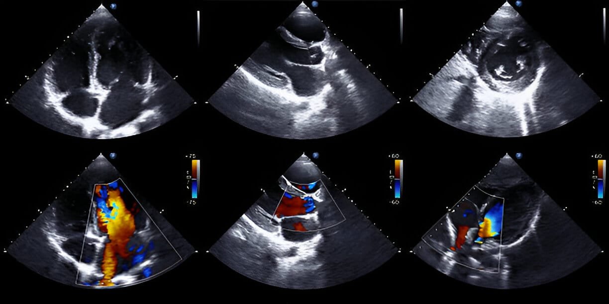
- Doppler Echocardiography: Measures the direction and speed of blood flow through the heart.
What is the Difference Between ECG and Echocardiography?
A common question patients have is about the difference between Echocardiography vs ECG. While both are crucial in cardiac diagnostics, they serve different purposes.
- ECG (Electrocardiogram): Records the electrical activity of the heart and helps in identifying irregular heartbeats, damage, or abnormalities in the heart’s structure.
- Echocardiography: Provides visual images of the heart’s structures and functions, such as the size and shape of the heart, its chambers, and valves.
While an ECG records the electrical impulses, echocardiography offers a visual snapshot of the heart’s architecture and motion.
What is Echocardiography Used For?
Echocardiography is a versatile tool used for various diagnostic purposes. Many patients wonder if an echo can detect blockage. While it is not primarily used for detecting coronary artery blockages, it can provide indirect evidence of such conditions by showing abnormal movement of the heart walls or other structural issues.
Echo Test Uses
- Diagnosing Heart Conditions: Evaluates heart murmurs, valve diseases, and congenital heart defects.
- Monitoring Heart Health: Assesses the effectiveness of treatments for heart conditions.
- Detecting Heart Muscle Issues: Helps in identifying cardiomyopathies and pericardial diseases.
- Evaluating Heart Function: Measures the ejection fraction, which indicates how well the heart is pumping blood.
What is a Normal Echo Report?
Understanding a normal echocardiography report can provide reassurance about heart health. A typical echocardiography reports normal values including normal chamber sizes, normal valve function, and an ejection fraction within the normal range.
Report Insights
- Chamber Sizes: Normal ranges for the size of the left and right ventricles and atria.
- Valve Function: Valves should open and close properly without significant leakage.
- Ejection Fraction: Normal values range from 50% to 75%, indicating the heart is pumping well.
How much time does an echo test take? Typically, an echo test lasts about 30 to 60 minutes. It is also asked if an echo can detect heart blockage. While it primarily evaluates structural issues, it can suggest blockages if abnormal wall motion is detected.
Echo test duration varies but generally falls within the 30 to 60-minute range.
About 2D Echo
A 2D Echo, or 2D echocardiography, is a widely used technique in cardiac imaging. The 2D echo test provides two-dimensional images of the heart, offering valuable insights into its functioning.
2D Echo Key Points
- 2D Echo Test: A 2D echo test is a sophisticated imaging technique that provides highly detailed images of the heart’s structure and function. By using sound waves, this non-invasive procedure captures moving pictures of the heart in real time, allowing cardiologists to assess various aspects such as chamber size, valve function, and overall cardiac performance with exceptional clarity and precision.
- What is 2D Echo: Also known as Two-Dimensional Echocardiography, a 2D echo utilises ultrasound technology to generate dynamic images of the heart’s anatomy. These images provide valuable insights into the heart’s chambers, walls, and valves, aiding in the diagnosis and monitoring of cardiac conditions. It is a vital tool in modern cardiology for its ability to visualise complex cardiac structures in great detail.
- 2D Echo Scan: The 2D echo scan is a safe and painless procedure conducted on the chest area. It involves placing a transducer emitting high-frequency sound waves over the chest, which then captures detailed images of the heart’s internal structures. This non-invasive method is crucial for diagnosing and evaluating various heart conditions without the need for surgery or invasive procedures.
- 2D Echocardiography Price: The cost of a 2D echocardiography test can vary depending on the healthcare facility and geographic location. At Atrius, we offer competitive pricing for our 2D echo services, ensuring accessible and high-quality cardiac imaging for all patients seeking comprehensive heart health assessments.
- 2D Echo Procedure: During a 2D echo procedure, patients lie comfortably on an examination table while a trained technician moves a transducer device over the chest area. The transducer emits and receives sound waves that bounce off the heart’s structures, producing real-time images displayed on a monitor. This procedure is painless and typically takes between 30 to 60 minutes to complete.
- 2D Echo with Colour Doppler: Adding colour Doppler to a 2D Echo enhances its diagnostic capabilities by visualising the direction and speed of blood flow within the heart and blood vessels. This advanced technique allows cardiologists to assess abnormalities such as valve regurgitation, blood clots, and congenital heart defects more accurately, contributing to comprehensive cardiac evaluations and treatment planning.
- 2D Echo Test Procedure: The 2D echo test procedure begins with the application of a gel on the chest area, which helps transmit sound waves efficiently between the transducer and the skin. The technician then moves the handheld probe (transducer) gently over the gel-coated skin, capturing detailed images of the heart from different angles. This straightforward procedure is crucial for evaluating cardiac structure and function in patients of all ages.
- How is 2D Echo Test Done: Similar to an ultrasound examination, the 2D Echo test uses a handheld probe that emits high-frequency sound waves. These waves penetrate the chest and bounce back (echo) from the heart’s structures, creating real-time images displayed on a monitor. This non-invasive method provides invaluable diagnostic information without exposing patients to radiation or discomfort.
- 2D Echo Uses: A 2D echo is instrumental in diagnosing a wide range of heart diseases, including coronary artery disease, heart valve abnormalities, cardiomyopathies, and congenital heart defects. It is also used to evaluate heart function, measure ejection fraction, and monitor the effectiveness of cardiac treatments. This versatile imaging technique plays a crucial role in guiding medical decisions and improving patient outcomes in cardiology practice.
Understanding the intricacies of electrophysiology and echocardiography can demystify the processes and prepare patients for what to expect. At Atrius, we offer comprehensive and advanced cardiac care, ensuring patients receive precise diagnoses and effective treatments.
Atrius: Your Trusted Heart Care Partner
When it comes to heart health, Atrius stands out as the best heart hospital in India. Our facility is equipped with state-of-the-art technology and staffed by the best cardiac surgeons in Delhi. Whether you need routine check-ups or advanced procedures, Atrius is your go-to heart hospital.
Why Choose Atrius?
- Best Heart Hospital in India: Our commitment to excellence has earned us this title.
- Heart Specialist Hospital: Focused solely on cardiac care, ensuring specialised treatment.
- Best Cardiac Surgeon in Delhi: Our surgeons are leaders in their field, offering world-class care.
- Best Bypass Surgery Hospital in India: We have a stellar record in performing successful bypass surgeries.
- Best Hospital for Bypass Surgery in India: Comprehensive care from diagnosis to recovery.
Schedule your consultation today with Atrius, the best heart hospital in India. Experience world-class care and advanced electrophysiology services designed to keep your heart healthy.
Visit Atrius or call us at +91 9818520234 to book an appointment now!
FAQs
Q: How do I prepare for an electrophysiology procedure?
A: Your doctor will provide specific instructions, but generally, you should avoid eating or drinking for a certain period before the procedure.
Q: Is echocardiography safe?
A: Yes, echocardiography is a non-invasive, safe procedure with no known risks.
Q: How long does it take to get the results of an echo test?
A: Results are typically available within a few hours to a day, depending on the urgency and facility.
For more information or to schedule an appointment, visit our website or contact us directly.
Atrius is committed to providing exceptional cardiac care and ensuring your heart health is in expert hands.