Treatments

Imaging Diagnostic Techniques in cardiology play a critical role in assessing the structure and function of the heart, identifying potential abnormalities, and guiding appropriate treatment strategies. Some commonly used diagnostic tests in cardiology are-
Electrocardiography is a non-invasive test that records the electrical activity of the heart over a period of time. It involves placing electrodes on the skin at specific points on the body, typically the chest, arms, and legs. These electrodes detect the electrical signals generated by the heart’s depolarisation and repolarisation, allowing for the creation of an electrocardiogram (ECG or EKG).
The ECG provides valuable information about the heart’s rhythm, rate, and electrical conduction pathways. The ECG waveform consists of several components, including the P wave (atrial depolarization), QRS complex (ventricular depolarization), and T wave (ventricular repolarization). Deviations from the normal ECG pattern can indicate various cardiac conditions, such as arrhythmias, heart attacks (myocardial infarctions), and electrolyte imbalances.
ECGs are widely used in various clinical settings, from routine check-ups to emergency situations. They can help diagnose and monitor conditions like atrial fibrillation, ventricular tachycardia, bradycardia, and heart blocks. ECGs are also valuable in assessing the effects of medications, detecting cardiac ischemia (lack of blood flow to the heart muscle), and evaluating the overall electrical activity of the heart.
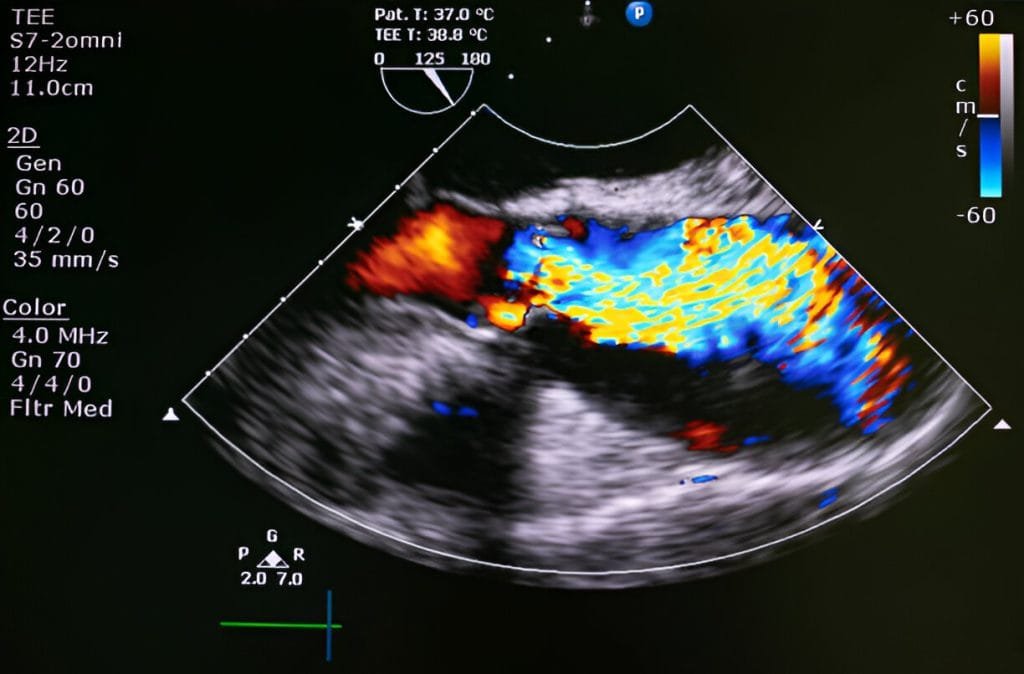
Echocardiography is a non-invasive imaging technique that uses sound waves to create detailed images of the heart’s structures, including its chambers, valves, and blood flow patterns.
During an echocardiogram, a transducer (a small handheld device) is placed on the chest or sometimes inside the oesophagus to emit high-frequency sound waves. These sound waves bounce off the heart’s structures and are captured by the transducer to create real-time images on a monitor. Echocardiography can be performed from different angles to obtain comprehensive views of the heart.
The technique provides critical information about the heart’s size, shape, function, and blood flow dynamics. It is used to diagnose and assess conditions such as heart valve abnormalities (stenosis, regurgitation), cardiomyopathies (enlarged or weakened heart muscle), congenital heart defects, pericardial effusions (fluid around the heart), and more.

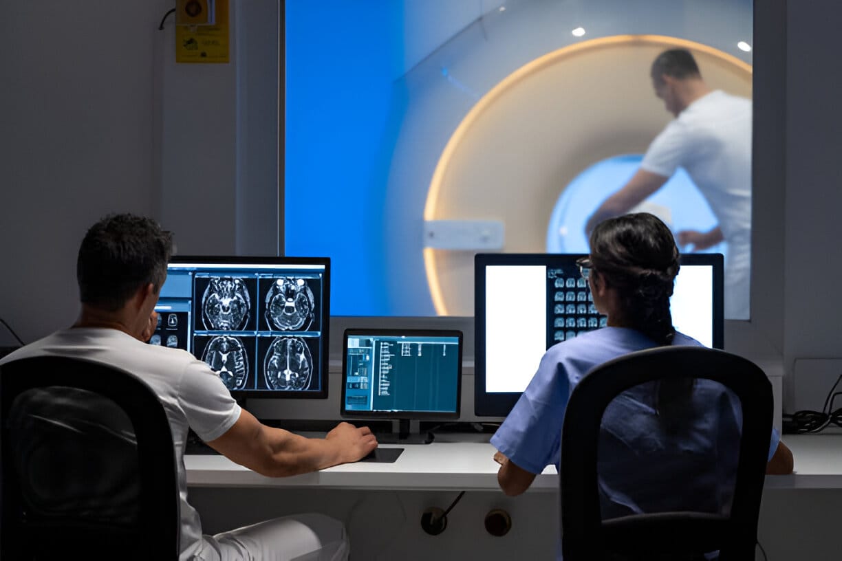
Cardiac MRI is a powerful imaging technique that uses a strong magnetic field and radiofrequency waves to create detailed images of the heart’s structures and functions. It provides comprehensive insights into both the anatomy and physiology of the heart.
During an MRI, the patient lies within a large machine that generates a magnetic field. Radiofrequency pulses are applied, causing the protons in the body’s tissues to emit signals. These signals are then captured by the MRI machine to create high-resolution images of the heart in multiple planes.
The test reveals the heart’s structures (chambers, valves, vessels), aiding in diagnosing defects, valve issues, and tumours. It evaluates function (ventricular volumes, ejection fraction), identifies tissue health, diagnosing conditions like myocarditis and scar tissue, and assesses blood flow and viability after ischemia.
Cardiac CT is a non-invasive imaging technique that uses X-rays and advanced computer processing to create detailed cross-sectional images of the heart and its blood vessels.
During a CT scan, the patient is positioned on a table that is moved through a doughnut-shaped machine. X-rays are directed through the body, and detectors on the opposite side of the machine capture the X-ray beams after they have passed through the body. Computer algorithms reconstruct these data into detailed images.
This is a highly versatile tool with multiple clinical applications. One crucial role is the assessment of coronary arteries, where it meticulously examines for calcifications, plaques, and stenoses. This helps diagnose coronary artery disease and aids in making informed treatment decisions.
Additionally, the test offers detailed visualisation of the heart’s chambers, valves, and other structures, facilitating the diagnosis of congenital heart defects and providing valuable insights for surgical planning. The technique’s ability to conduct calcium scoring further enhances its value by estimating the risk of coronary artery disease through quantifying calcified plaque in coronary arteries.
Contrast agents are pivotal in both cardiac MRI and CT, enhancing image quality and highlighting specific anatomical features or blood vessels. They are typically administered intravenously, contributing to the precision and effectiveness of these imaging modalities.
These advanced diagnostic tools play an essential role in diagnosing various cardiovascular conditions, guiding treatment decisions, and monitoring disease progression- forming a comprehensive toolkit for cardiologists to assess heart health and provide personalised patient care.